CT PricingWe would be happy to serve your clients during daytime hours!
*The radiologist report gets e-mailed directly to you! Two ways to refer:
|
Prices:
**All prices include anesthesia, IV catheter, cardiopulmonary monitoring, and radiologist report 1. Dental/skull/sinuses: $350 2. CT one region: $500 3. CT two regions: $600 4. CT with contrast: (Myelograms) <20 lbs = $650 20-40 lbs = $750 >40 lbs = $850 |
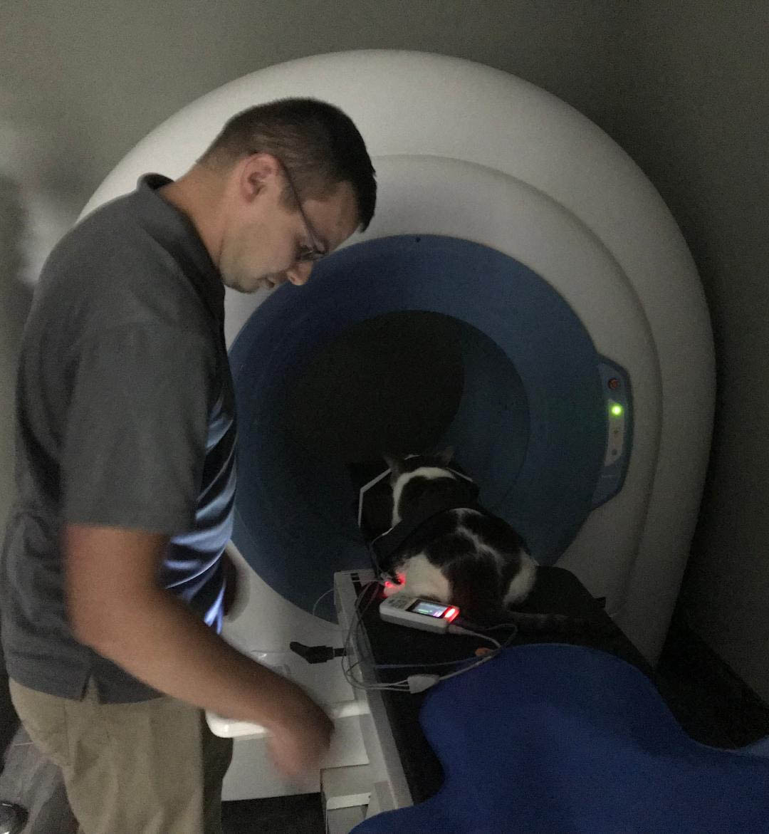
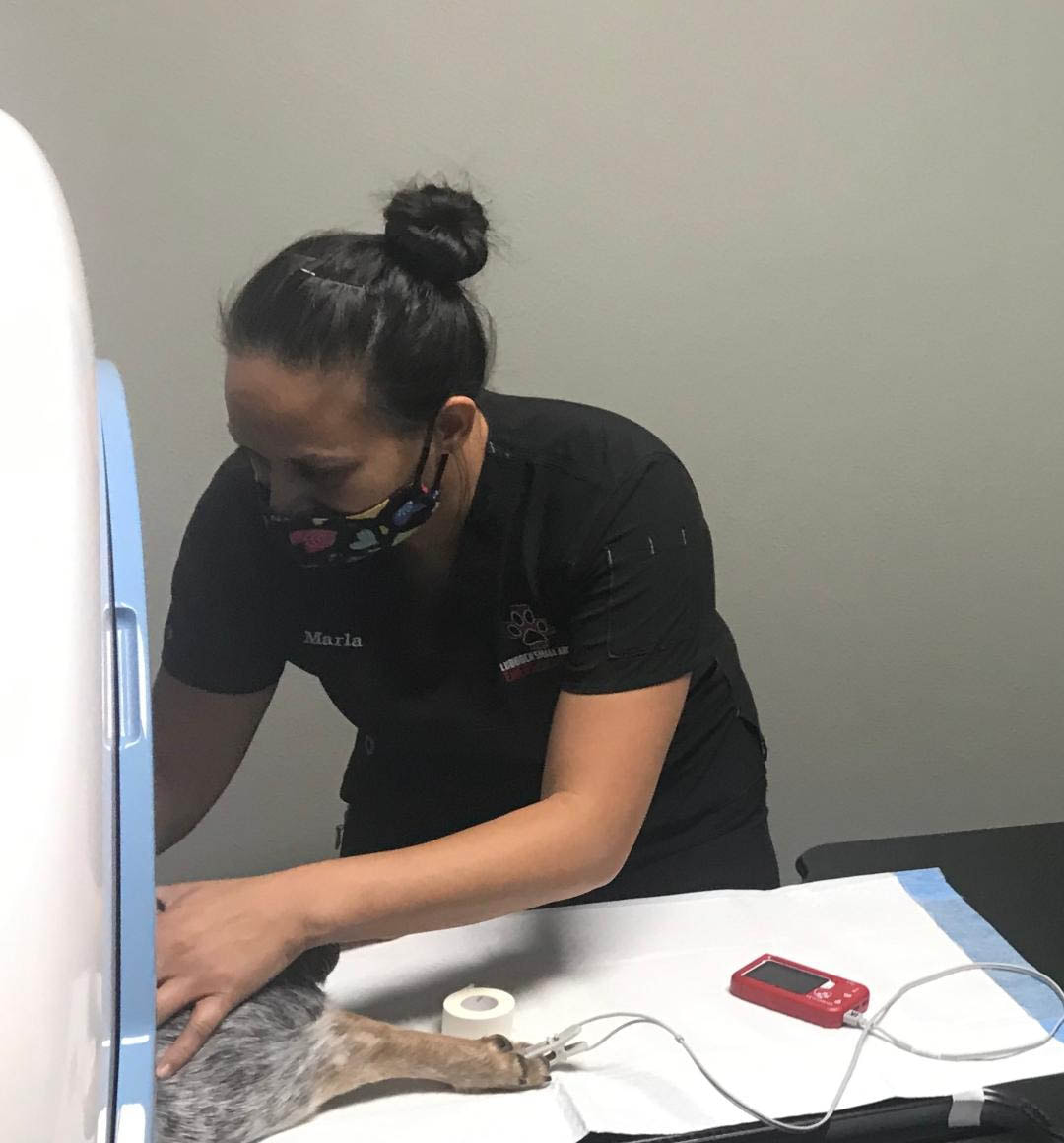
Lubbock Small Animal Emergency Clinic offers CT imaging for all your patient’s needs. |
CT scans are particularly useful in detecting issues, such as:
When can I schedule a CT scan for my patient?
We offer CT scans every day of the week but most of them are performed Monday through Friday. We will set up a time that best works for the client to drop off the patient. To schedule a CT scan:Call and leave a message at the clinic at 806-797-6483 |
The date and time for your patient’s CT scan has been set. What is the next step?
|
How can my pet benefit from a CT Scan?Axial Skeleton Bulla studies. CT is the modality of choice for differentiating infectious, inflammatory and neoplastic processes involving the ear. It can clearly delineate between otitis externa, otitis media, and neoplasia. It defines the extent of bone involvement, i.e. reactive otitis, osteolysis.
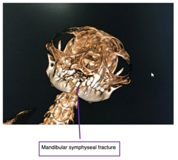
Nasal and Sinus Studies: CT is superior to radiographs in defining destructive from non-destructive rhinitis, sinusitis and neoplasia, as well as the extent of involvement, and location. Also useful in the localization of foreign bodies and tooth root abscesses. Skull Hemorrhage: CT is the modality of choice for recent intracranial hemorrhage. There is a linear relationship between the amount of hemoglobin (Fe content) and x-ray beam attenuation. The hemoglobin content is highest within 24 hours of a bleed. Potential causes include trauma, DIC, neoplasia, anticoagulants (rodenticide toxicity), infectious (fungal granuloma formation with vascular erosion). Trauma/Fractures: CT is excellent at defining the type and extent of fractures, as well as the extent of adjacent soft tissue involvement. This is important in determining prognosis, management (medical vs. surgical), and surgical planning. Appendicular Skeleton Elbows Dysplasia: CT is superior to radiographs in defining lesions of the elbow. A CT scan is warranted in all young dogs with a history of persistent lameness localized to the elbow, especially if radiographs are normal. At most institutions, an elbow CT study is part of the routine protocol in all animals meeting these criteria. Lesions commonly diagnosed include fragmented medial coronoid process, ununited anconeal process, OCD, DJD, joint incongruency and joint surface defects Ectopic Ureters: Contrast CT is excellent for differentiating between unilateral vs. bilateral, intramural vs. extramural, and location of termination. This information is helpful in determining surgical approach; which in return reduces surgical time. |
Trauma:
CT is superior to radiographs in defining small, non-displaced fractures (i.e. fissure fractures, cortical stress fractures), especially in the distal limbs (carpus, tarsus). In certain instances of acute trauma, initial radiographs may be unrewarding and would require 7-10 days before the fracture was visualized. In this situation, because of its superior contrast resolution and cross-sectional capabilities, a CT of the effected bone would be warranted. CT also provided useful information regarding soft tissue/muscle trauma and is considered a viable option in debilitated, geriatric and potential anesthetic risk patients because of the rapid scan time or when cost maybe an issue. Abdomen Portosystemic Shunts: Contrast CT is a non-invasive, sensitive and specific means of identifying, localizing and determining the number of both intra and extrahepatic portosystemic shunts. Pancreatitis: The use of contrast CT in evaluating for pancreatitis is very sensitive in defining the effected region, the viability of the effected region, and whether surgery is indicated. CT is sensitive in differentiating between necrotizing and non-necrotizing pancreatitis. Mid abdominal masses: Based on tissue kinetics and the vasculature supply of different lesions, contrast CT is becoming increasingly accurate at differentiating between neoplastic, infectious and inflammatory processes. CT is also helpful in defining location, extent of involvement and surgical vs. non-surgical masses. Thorax Pulmonary Metastases: CT is considered the gold standard when evaluating for pulmonary metastases. Other applications: Pulmonary thromboembolic disease (PTE), chylothorax, pulmonary contusions, fractures, bronchial or upper airway involvement, and chronic lower airway disease. |


